Level 1 of DMS is coming to an end, I thought I would share what I found was most interesting. There were many things in every class – I could not write about it all otherwise this post would be too huge.
Physics

- The Ultrasound gel that is used is not only for preventing friction, it acts as a medium to help the sound waves move into issue, without it, all the ultrasound waves would reflect away.
- We have all heard about megabytes, but did you know there is such a thing as a nibble? 1 Megabyte = 2 Million nibbles
- A Piezoelectric element is what make US pulses, when you apply electricity it puts pressure on the piezoelectric element creating vibrations. The element not only sends out ultrasounds but also “listens” to the sound waves coming back. Bones can act as a piezoelectric material.*
- Imagine how fast the ultrasound beam has to be to travel deep into the tissue and back again several times a second to create a whole continuous image. It sends a pulse then listens and repeats, over and over again.
- The speed of sound through soft tissue is ~1540m/s, I found on google that the fastest car is 270.49mph which equals 120.9m/s. Diagnostic US frequency is at 1MHz-20MHz, we can only hear 20 to 20,000 Hz.*

Cardiac
- Most people experience premature ventricular contraction, it is where your ventricle contracts early before it had time to fill with blood. It feels like a skipped beat, even though you had an extra beat. Since starting this program I have gotten really good at noticing when I get them, one causation is stress.
- I was surprised to learn that many people have regurgitation in their heart valves, it’s not significant. We even use the tricuspid regurgitation to calculate pulmonary pressure. If you don’t have it, we can’t calculate it.
- When watching the mitral valve it looks like it flutters shut or flaps twice in one beat. This is because it opens and passively fills the ventricle, it starts to close but then a second “kick” opens it wide and the rest of the blood from the atrium shoots into the ventricle.
- We image the heart upside down, we aren’t upside down but the heart is.
- A heart attack is pretty specific to the coronary arteries that surround the heart and how it is not supplying the heart muscle with adequate oxygen and removal of waste (due to a blockage, for example).
- I was unaware that they do a transesophageal echo often for heart surgery or to show blood clots before defibrillation someone. Grey’s Anatomy never taught me that.
Abdomen

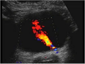
- You can see jets of urine filling the bladder through the ureters.
- When you lay on your side your gallbladder flops over, it can move towards your great vessels.
- Some people have an accessory spleen, that isn’t attached to the OG spleen. If you remove your spleen the accessory spleen can grow and take over.*
- If your ligaments loses laxity in the spleen, you can get what is called a wandering spleen.*
- You can live without a gallbladder because the bile will sit in your bile ducts, but you cannot store as much.
- We can tell if you’ve eaten a fatty meal, because your gallbladder contracts.
- We learned in high school that the large intestines does the majority of the absorption, but actually the small intestines does.
- Seminal vesicles can look like a bow-tie or a mustache on an ultrasound.
Women’s Health

- From looking at an ultrasound of the uterus you can tell at what stage in the menstrual cycle a women is at. You cannot see the exact point of ovulation but you can tell they are in that stage.*
- Ovaries are not fixed in one position, you have to look for them. Sometimes they are hiding behind bowel, sometimes one is sitting on the uterus, sometimes one is high and one is low. It’s an Easter egg hunt.
- Endometriosis is where you have endometrial tissue, that is usually found in the uterus, can pop up somewhere else in the body (but usually the ovary). Sometimes called chocolate cyst, it can bleed too when your going through your “cyclic regeneration”.*
- Dermoid cysts are a benign mass found in ovary, they could have teeth, hair, brain matter, etc… Do not google it!
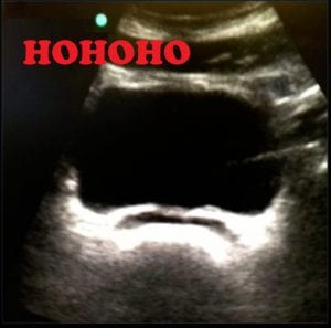
- When bladder is empty your uterus usually flops forward. Kind of like a deflated balloon.
*Thank you to some of my classmates for their input on what they thought was interesting.
Enjoy the winter break everyone, good job, we did it, we finished Level 1! Good luck to the Level 3’s who are off to Clinical in January, thank you for all your help. It will be weird without you guys next term.

