It’s that time again, level 2 of DMS has ended, here is a list of things I found most interesting. This was a long semester, from January to May, but full of tons of information and interesting facts. This is part 1 of 2.
Vascular

- A carotid study is looking for narrowing in the vessel of the neck caused by plaque. If a piece of this plaque breaks off it can cause a stroke.
- A deep vein thrombosis study is to rule out blood clots in the leg. DVTs can cause a pulmonary embolism, which can be deadly.
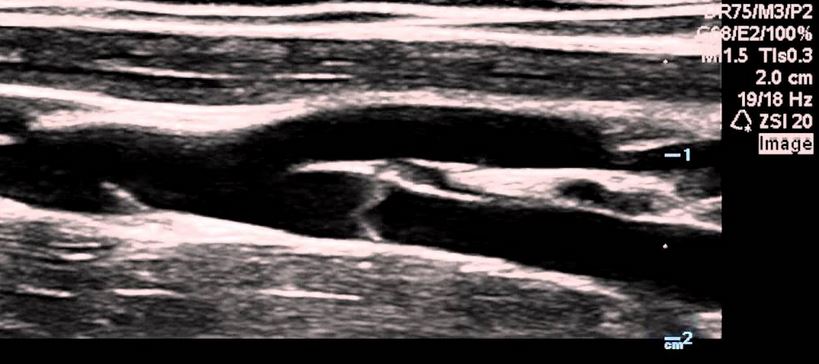
- You can see the valves in veins! Veins need more help sending blood back to the heart. These one way valves help blood move forward and prevent them from moving backwards.
- The muscles in your legs help move the blood from your feet up to your heart. If you stand too long, blood will pool and not be able to make its way back and you could faint.
- It is not uncommon to see duplicated leg veins (some people have quadrupled leg veins). Many have a variant called giacomini vein. It is an extension of a vein in the calf, instead of terminating it continues. I have that.

- To help prevent DVTs wear compression socks on long flights or make sure to move around. Oral contraceptive pills also increase your risk of DVTs.
- The common carotid artery supplies to our brain with nutrients and oxygen. It has low resistance flow which means that we have a constant blood supply to our brain.
- Homonymous Hemianopia: if you were to split your eye into 2 halves, this condition causes you to only see through one half, either the left half or right half in both eyes.

eg of Homonymous Hemianopia - We can tell the difference between internal carotid artery and the external carotid artery just by listening to it using Ultrasound.
- We can hear pathology. If there is a narrowing of a vessel and we were to ‘listen’, you can hear it.
Cardiac
- A big role of what we do as ultrasound echotechs is to evaluate systolic (how well the heart functions) and diastolic (how well the heart relaxes) function of each patient.
- Systolic function goes down when the heart wall gets damages from a heart attack or if the ventricle stretches too much from an increase in blood volume.
- Diastolic function decreases when the heart muscles gets too thick which is caused by increase in pressure in the heart. As we age we all have some diastolic dysfunction. It has a higher risk of morbidity and mortality.
- When the heart relaxes it is an active process. This means that work needs to be done to help pump the heart and to help it relax. The heart takes no breaks.
- In a heart transplant surgeons divide the patients left atrium, leaving the back wall with the pulmonary vein and attaches the new donor heart. I thought they take the whole heart and replace it with the new one.

- The most common cause of sudden death in young athletes is Hypertrophic Cardiomyopathy. It affects 1:500 people and is an autosomal dominant trait. If one of your parents has this you have a 50% chance of also having HCM. The heart muscle thickens and has difficulty relaxing. The lethal arrhythmia associated with HCM causes sudden cardiac death.
- High Blood Pressure: if not treated causes your heart muscles to enlarge which can cause diastolic dysfunction. 1:6 Canadians are unaware they have high BP. They call this the silent killer.
- After checking every patient for systolic and diastolic dysfunction if there is additional issues discovered, we have to tailor our exam and perform additional procedures depending on what it is.
Photo cred: featured image, vein, jugular, HH, cat, and heart drum
Content cred: DSON2000 level classes


