When I talk about the Medical Diagnostic Sonography program these are some of the reactions I get:
1. Confusion
I understand that people get confused due to being more familiar with the term Ultrasound. I can admit I didn’t know what Sonography meant at first either.
2. “Did you say Stenography?”
A stenographer, according to Google Dictionary is “a person whose job is to transcribe speech in shorthand.” No, I am not a Stenographer.
3. “Oh, so you ultrasound babies”
We may do obstetrics but it is only a portion of what we learn and what we are trained to do.
From what I have learned so far this is what people should know about Sonographers:
1. We have a wide scope of practice and can perform ultrasounds on the abdomen, superficial structures, pelvis, breasts, pregnancy, carotids, peripheral veins and heart. We do diagnostics, contrast studies, research, therapies and treatments.

2. Air is not a Sonographers friend, we cannot see through the lungs or the Gastrointestinal tract. GI gases frequently gets in the way for imaging organs. If bones are in the path of the beam, we have to figure out ways around them because we cannot see through them.
3. Sometimes we correlate our findings using other imaging modalities such as MRI, CT, nuclear med and X-Ray. With the help with Med Lab techs, we can piece together the possible reasons a patient is sick. For example, if a patient has elevated bilirubin this might lead us to check to see if there is an obstruction in their bile duct.
4. Sometimes we give Surgeons a road map to guide their surgeries by telling them a mass is located. We assist doctors with ultrasound guided biopsies, as the needle enters the body we use the ultrasound as a ‘camera’ to tell the doctor where the needle is and where they need to go.
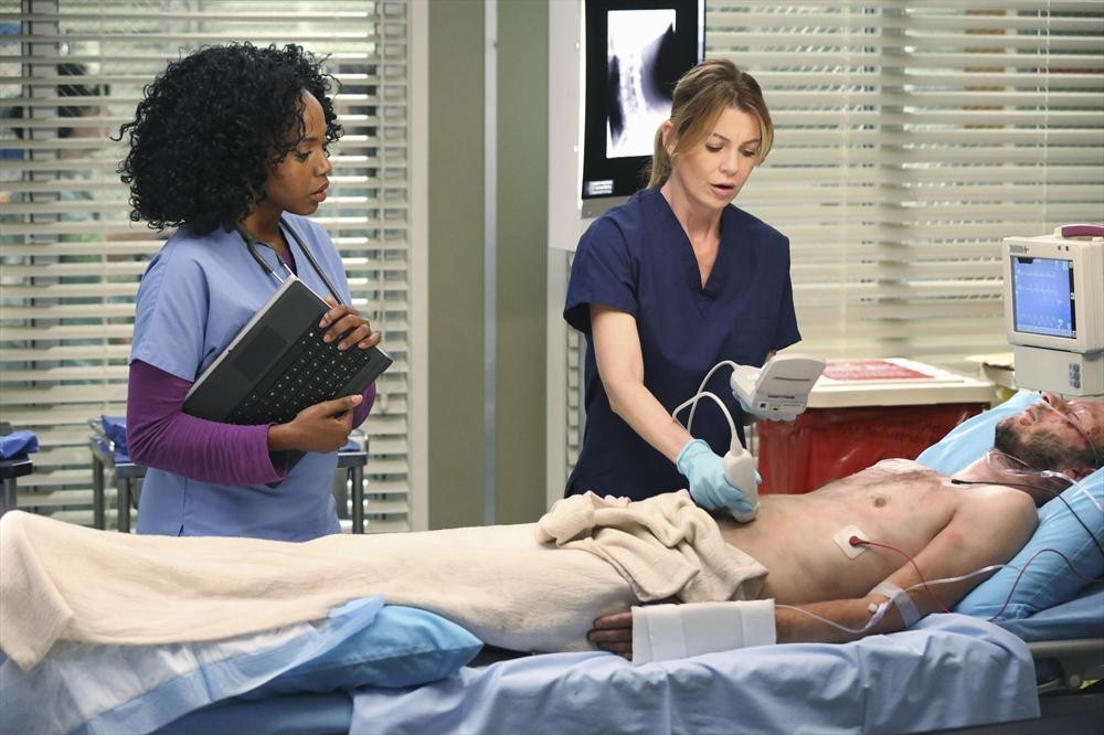
 5. In TV shows the doctors perform the ultrasounds. From my understanding, doctors rarely perform US, only in certain cases.
6. We perform measurements: kidney sizes, velocities of blood, cross-sectional areas of heart valves, fetal abdominal circumference, volume of blood ejected from the heart, etc… just to name a few.
7. We need to be able to identify pathologies because patients and doctors rely on Sonographers on their expertise. We report our findings to the radiologist and we have to do continued education while working.
8. We perform a full abdominal routine where we will scan the aorta, IVC, liver, pancreas, gallbladder, and kidneys. Even if you were just in for gallstones. We have to learn several different routines depending on what the doctor is looking for.

9. We have different probes for different tasks. To make sure that we have the best diagnostic images, we constantly have to change out technical settings. Gain (brightness), Frequency (lower frequency for visualizing deeper organs and higher frequency for visualizing shallow ones), depth and focus (clears up specific area of interest) are some of them.
10. We have to control patients breathing and position. We sometimes require that the patient have a full bladder or not eat hours before the exam. All these things are important to get the best image possible for diagnostic use.
Photo cred: DMS, alien US, Stomach, Grey’s Anatomy and US wands

