Happy December, folks! I am now into the third department of my rotation—Hematology, which is the study of blood and blood disorders.
There are three categories of blood cells: white blood cells (WBCs), red blood cells (RBCs) and platelets. All three arise from stem cells in the bone marrow, differentiating to perform diverse roles in the human body. WBCs defend against infection and other foreign materials. RBCs – the most abundant cell type in the blood – carry oxygen from the lungs to the tissues. Platelets are responsible for primary hemostasis, the first step of blood clot formation.

A complete blood count (CBC) is a common blood panel performed on a hematology analyzer. The test measures several WBC, RBC and platelet parameters, therefore providing a comprehensive picture of all three cell types’ counts and morphologies. If a result flags as abnormal or the analyzer is unable to differentiate the cells, a manual blood film examination may be ordered. Physicians can also order a manual blood film or pathologist review, regardless of the automated results, if a clinical condition is suspected or being monitored.
I’ve spent the past couple weeks on the ‘diff’ bench, which is where blood films are made, stained, and read under the microscope. There are three major constituents of a blood film examination and depending on the patient’s CBC results and clinical history, zero to all three are performed.
Platelet estimate – May be ordered to verify an abnormally low, and to a lesser extent high, automated platelet count.
RBC morphology – Assesses cell size, variability in size, cell shape, colour, and the presence (or absence) of cellular inclusions.
WBC differential – A differential involves manually counting and classifying 100 WBCs to determine their percent distribution. The five types of white cells are neutrophils, lymphocytes, monocytes, eosinophils and basophils, with immature and abnormal forms denoted separately.
Be the first to receive the latest BCIT News. Become a subscriber.
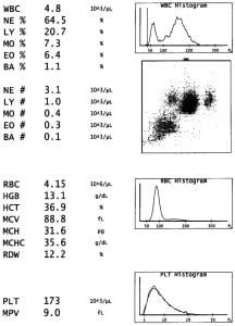
Here is a typical CBC analyzer printout. The WBC indices are a total count (4.8 x 10⁹/L ), and the counts of the 5 WBC types as a relative percentage and absolute value. There are 7 RBC indices on the printout, and 2 for platelets. In addition to numerical values there is a scatter plot separating white cells by size and granularity, and histograms showing WBC, RBC, and platelet-size distribution curves – all of which are normal on this printout.

We had lots of practice reading blood films, a.k.a. peripheral blood smears, at BCIT; the part I found most challenging was correlating a patient’s abnormal findings with potential causes; narrowing the list and following that train of thought until the results are verified.
I’ll do a mini case study in my next blog, but essentially, there is more critical thinking and follow through involved with hematology specimens in comparison to chemistry specimens. Even if a sample is flagged on a chemistry analyzer, and you have to manually review the results or dilute it—once that is done, you’re done with the sample. In Hematology, there is more investigation into a patient’s clinical history; deciding what does, and does not, require a peripheral blood smear or referral to a pathologist.
Learn more about the Medical Laboratory Science full-time diploma program at BCIT.
